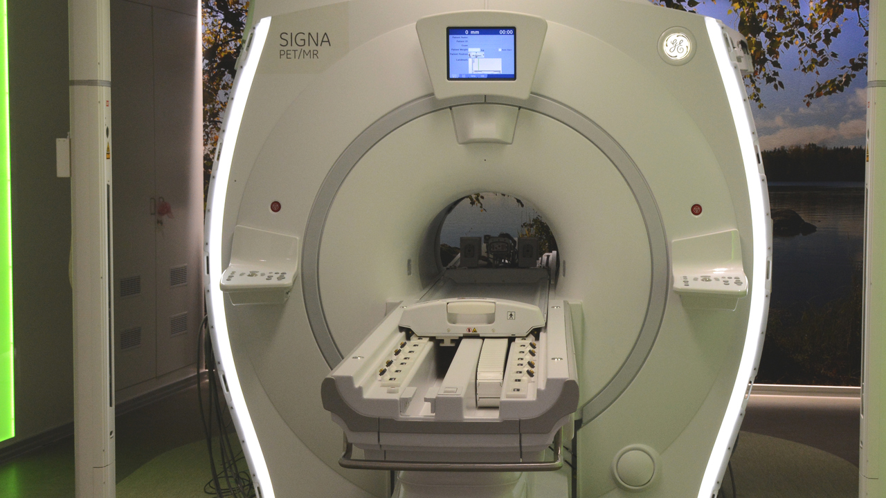
Magnetic resonance imaging (MRI) offers great potential when it comes to diagnosing cancer. However, the scanning procedure is very sensitive. One of the many problems can be that if the patient moves slightly during the examination, the images become blurry. This makes it more difficult to accurately determine the size and location of a cancerous tumor. It is important to be able to plan treatment precisely, for example, directing radiation so that it attacks the tumor but not the healthy tissue surrounding the tumor.
On the other hand, as medical technology improves, the amount of data available to help treat a patient increases, but it also greatly increases the workload of already overburdened medical staff. It can create information pressure that can cause delays and errors. More efficient image processing operations would therefore be a much-needed improvement.
In his thesis at the Department of Radiation Sciences, Attila Simko demonstrates how to improve the quality and efficient processing of MRI images using machine learning. In the studies presented, Attila and colleagues developed machine learning models that were trained to remove common artifacts in MRI images, including noise and motion. They have also developed a powerful model for creating synthetic CT scans from MRIs. To further develop their methods, they are all made publicly available for researchers to further use and compare.
Attila Simko created a web version of his thesis with several interactive characters to help understand the field. More if it is running www.mlsatellite.com/kappa.

“Extreme tv maven. Beer fanatic. Friendly bacon fan. Communicator. Wannabe travel expert.”


More Stories
Why Rare Earth Metals for Electric Cars Are Crucial for Modern Mobility
“We want to promote critical rules approach”
“A lot happened during the trip,” Jönköping County Council