An orthopedic prosthesis means a very different quality of life than a traditional prosthesis for those who have had to have a bone amputation, for example. Currently, the number of patients who can receive the implant is limited by the fact that they must have a good ability to heal. Professor Anders Palmquist studies what happens at the meeting between the implant surface and patients’ bone tissue. The purpose is to allow a much larger number of amputees to receive an implant. An advanced microscope, funded with funds from the Lundberg Research Foundation, will contribute to new nanometer-level knowledge about how bone heals and develops around an implant.
Traditional dentures often cause problems for their users. The fit is rarely perfect, they are uncomfortable to sit on and the skin becomes sweaty and irritated in the arc between the sleeve and body. An orthopedic prosthesis is attached with titanium screws (implant) to the patient’s skeleton. Studies show that patients with orthopedic prostheses use their prostheses more, increase their mobility, reduce problems and improve quality of life.
Most amputations in Sweden are performed due to diabetes. Currently, diabetics cannot get an artificial implant because diabetes also means a poor ability to heal. The same is true for patients with osteoporosis and cancer patients treated with radiation. Therefore, more knowledge is needed about the bone tissue of these patients and its ability to heal and grow.
Bone cells that communicate
Anders Palmquist, Professor in the Department of Biomaterials Sciences at the Sahlgrenska Academy at the University of Gothenburg, focuses his research on how bone tissue forms around the implant. The goal is to enable more people to be treated with orthopedic prosthetics. He has developed new methods for analyzing the chemistry and structure of bone tissue that make it possible to see how diseases, lifestyles, and medications affect bone quality and growth. It’s about causing dizziness up close under the microscope. At the nanometer level, bone tissue looks like a mat made up of several layers of cords, which in turn are made up of many tiny strands that are proteins with minerals around them. They are the ones who make bone strength.
By analyzing the structure of the bone, we can see how it was formed. We’re looking at osteoblasts, the cells in bone tissue that feel the load, and the connecting filaments that are between 200 and 300 nanometers in diameter. They have direct contact with the implant surface and signal to the body’s system to activate other cell types needed for bone formation and remodeling. “
Effects of pregnancy on bone growth
Thanks to a SEK 3 million grant from the Lundberg Research Foundation, Anders Palmquist can now further develop the analytics. Money finances the purchase of an AFM microscope*. This is often used to measure surface topography, for example in a tissue sample. But Anders Palmquist sees other possibilities.
The microscope has a very thin tip that measures just above the surface. We want to squeeze it into the material and measure the forces, the stiffness, in the tissue. Then we can relate bone structure and chemical composition to mechanical properties and understand more about how bone is affected by both implants, different disease states and how it is loaded. When walking or standing on an orthopedic prosthesis, the load on the skeleton rises. The skeletal bone tissue responds to the load and changes all the time to create the maximum strength based on the load it is exposed to. We need more knowledge about this process.”
New possibilities with simulation
Together with fellow researchers in Israel, Anders Palmquist is also working to build a knowledge bank of all the data that emerges. The knowledge bank will be a platform to simulate bone growth around implants.
“By using simulations, we can continue to build new knowledge while at the same time reducing experimental research. For example, we can assume that bones are forming at a certain speed and see how speed affects the mechanical development of bone. Then we can add additional parameters and see their effect on bone tissue.”
Another important area to learn more about is how the implant surface interacts with biology. Via doctoral student Paula Giraldo Osorno, Anders Palmquist’s research group is participating in an EU project studying how to alter the surface of implants so that cells in bone tissue thrive and grow well while bacteria do not.
“Infection is a big problem and we don’t want to have to use antibiotics,” Anders Palmquist explains.
More individual adaptation
And Anders Palmquist’s research should not just allow more people to get implants. The implants will also be improved through individual adjustment.
“With the possibilities in simulation, we will be able to develop ways to tailor the materials and surface properties of the implants specifically to each patient’s bone type and thus obtain a well-functioning localized healing. In addition, the implant design can be further modified to suit each patient.”
The new microscope has not yet been placed, but the buying process has begun.
“Different AFM equipment is good at different things. We want a microscope that can be used by many colleagues here in the department, but it has to be really good for our needs.”
* AFM: Atomic force microscope
For more information please contact:
Christina Bachmann
Chairman of Board of Directors
Lundberg Research Foundation
Mobile: +46727 19 70 45
[email protected]
first larco
Member of the Board of Directors
Lundberg Research Foundation
Mobile phone: +46734 33 71 40
[email protected]
Anders Palmquist
Mr
Department of Biomaterials Sciences,
Sahlgrenska Academy, University of Gothenburg
Mobile: +46706 83 29 66
[email protected]
Photographer: Magnus Gotander
Captions:
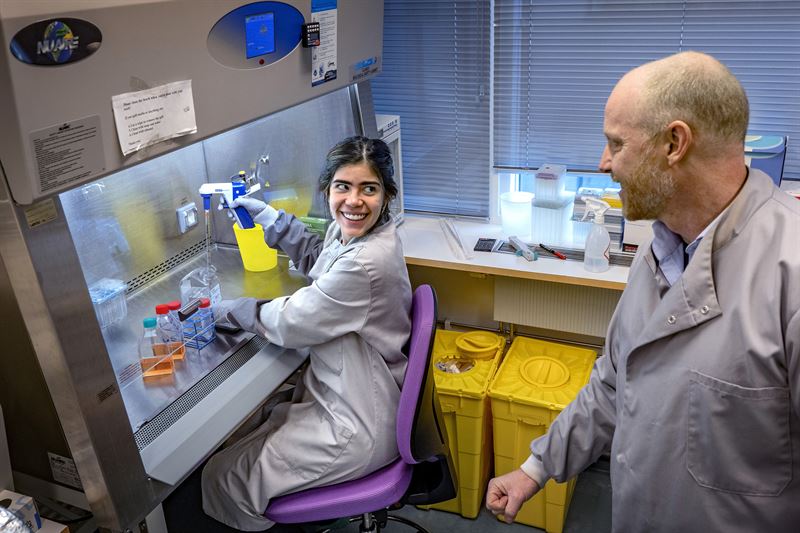
- PhD student Paula Giraldo Osorno and Professor Anders Palmquist in the lab
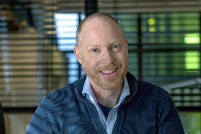
- Professor Anders Palmquist
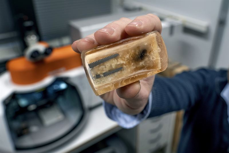
- Implanted in bone tissue
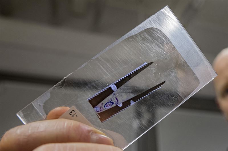
- planting
IngaBritt and Arne Lundbergs Forskningsstiftelse, which celebrates this year its fortieth anniversary, It was founded by IngaBritt Lundberg in 1982 in memory of her wholesaler husband, Arne Lundberg, born in 1910 in Gothenburg. The foundation aims to promote medical scientific research mainly related to cancer, kidney disease and orthopedics and to give priority to the procurement of equipment, aids and equipment. During the years 1983 to 2021, 573 grants were awarded with a total value of SEK 965 million, of which SEK 37 million was in 2021. Research in the Gothenburg region takes precedence. The headquarters of the Foundation is located in Gothenburg.
www.lundbergsstiftelsen.se
Anders Palmquist About his research:
Orthopedic amputated prostheses improve the physical function and quality of life of patients. By developing new methods based on nuclear energy microscopy, we are exploring how mechanistic development of bone tissue occurs at the nanoscale and how it is affected by implant design, pregnancy, and disease. The knowledge is used to improve implant design to treat patients with reduced skeletal quality and healing capacity.”
Tags:

“Extreme tv maven. Beer fanatic. Friendly bacon fan. Communicator. Wannabe travel expert.”







More Stories
Why Rare Earth Metals for Electric Cars Are Crucial for Modern Mobility
“We want to promote critical rules approach”
“A lot happened during the trip,” Jönköping County Council