The findings are a step on the way to finding better and more sophisticated tools to support clinicians who have to assess the thickness of melanoma, which in turn influences clinical decisions and priorities for care, says Sam Polisi, a researcher at the Sahlgrenska Academy in Gothenburg.
When diagnosing melanoma, dermatologists evaluate whether it is an aggressive form, in which cancer cells grow in the dermis and risk spreading, called invasive melanoma, or a milder form that only grows in the top layer of the skin, called melanoma in situ, MIS.
Invasive melanomas that grow more than one millimeter deep in the skin are considered thick and therefore also more aggressive.
Skin cancer facts
Melanoma is the most dangerous form of skin cancer and the cancer is on the rise. Melanoma occurs when the melanocytes in the skin begin to divide uncontrollably. Melanoma grows, often has several colors and generally has an irregular shape. The change may look like a new black brown spot or nodule on the skin, or a birthmark that changes. Often, melanoma also stands out in comparison with other birthmarks of the patient.
Thickness provides information about the prognosis
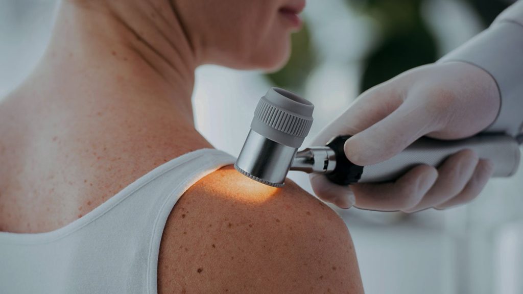
Melanoma is evaluated with a dermatoscope, which is a type of magnifying lens equipped with a strong light. Diagnosing melanoma is often relatively easy, but estimating the extent of melanoma is a much greater challenge.
— In addition to thickness, which provides valuable predictive information, it can influence the choice of surgery margins for the first operation and how quickly the operation is performed, says Sam Polisi.
Via a web platform, 438 international dermatologists evaluated 1,456 images of melanoma taken using a dermatoscope. The dermatologists’ results were then compared to those of a machine learning algorithm trained to classify the depth of melanoma.
Equally between the doctor and the machine
Dermatologists as a group had an average accuracy rate for correctly classifying melanoma depth of 63 percent for milder melanomas and 71 percent for invasive melanomas. Doctors were able to correctly determine that the melanomas were invasive or MIS in 76 percent of all evaluations. The corresponding percentage for the leading algorithm was 74 percent.
Dermatologist’s joint assessment of pre-trained algorithmic level in differentiating between MIS and invasive melanoma. Interestingly, the doctors’ professional background and experience with dermatoscopy had no effect on the diagnostic accuracy of predicting melanoma thickness, says Sam Polisi.
AI can become a valuable aid in healthcare
The development of artificial intelligence, AI, is advancing in many areas in healthcare. In particular, it is expected that this technology will be able to develop and become supportive of medical imaging, that is, for clinicians who evaluate and interpret images, for example x-rays, retinal images and skin changes. The technology can also be applied in areas other than image recognition.
Our study notes the difficulties in correctly assessing the thickness of melanoma based on dermatoscopy images. In future studies, we aim to examine the utility of pre-selected dermatoscopy structures for differentiating between thin and thickened melanomas. We also want to test whether clinical decision-making can be improved in this case using machine learning algorithms, says Sam Polesie.
The study was conducted in collaboration with researchers at the Medical University of Vienna in Austria.
Scientific study:
Assessment of melanoma thickness based on dermatoscopy images: an open web-based international diagnostic studyJournal of the European Academy of Dermatology and Venereology
Contact:
Sam Polesie, Lecturer at the Sahlgrenska Academy at the University of Gothenburg and a dermatologist at the Sahlgrenska University Hospital, [email protected]

“Extreme tv maven. Beer fanatic. Friendly bacon fan. Communicator. Wannabe travel expert.”


More Stories
Why Rare Earth Metals for Electric Cars Are Crucial for Modern Mobility
“We want to promote critical rules approach”
“A lot happened during the trip,” Jönköping County Council