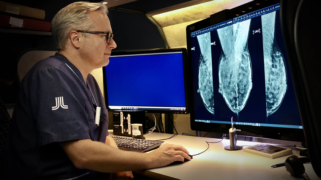
Fredrik Strand is driven by making a difference. That’s why he became interested in his field of research: how artificial intelligence could be used to review mammograms and detect suspected cancer. Early detection of cancerous tumors is important to be able to start treatment quickly and thus save lives.
– It started when she became interested in the field of breast cancer. He says: Because breast cancer is such a common disease, we can help many people by improving breast cancer diagnosis.
Fredrik Strand has been involved with computers and programming since childhood. Even as a 10-year-old, he was fascinated by the possibilities opened up by computers. When, many years later, he chose a research direction, artificial intelligence became a way to combine his education and areas of interest.
– Even when I was studying technical physics in Lund, I was fascinated by artificial intelligence. He says there was something magical about how computers mimicked human behavior.
Collaborates with KTH
Today, Frederik Strand divides his time between Karolinska University Hospital, where he works as a radiologist, and Karolinska Institutet, where he is an associate professor. His research group, which also collaborates with KTH, is developing, validating and testing artificial intelligence algorithms that will detect cancer.
The human mind has a limited size, but artificial intelligence does not.
In traditional mammography screenings, each mammogram is reviewed by two radiologists to evaluate whether the person needs to call back for further examination. In a recent study, ScreenTrust CAD, published in the journal Lancet Digital Health in September, Fredrik Strand and his research team were able to show that the tested AI algorithm and a radiologist were better than two radiologists at detecting suspected cancer in images.
– In several previous studies, and then on archival images, it was possible to prove that the latest generation of artificial intelligence was as accurate as an X-ray doctor in detecting cancer. But this does not automatically mean that it will work in everyday clinical practice. Fredrik Strand says: In our new study, we were able for the first time to prove with statistical certainty that artificial intelligence with a radiologist contributed to the detection of more cancers during screening.

In another study, ScreenTrust MRI, the aim was to evaluate women after regular mammography who needed further screening with magnetic resonance imaging (MRI). MRI is a more sensitive screening method and also picks up cancers that are difficult to detect on regular mammograms for various reasons.
To identify these women, it turned out that the AI algorithm was not enough, but the researchers had to combine three different types of algorithms that look at different parameters in the images. The algorithms were developed in collaboration with researchers from KTH.
“Algorithms must be trained to distinguish between images with and without breast cancer.”
It is exciting, but also challenging, to collaborate in this way across different academic disciplines, according to Fredrik Strand.
-It takes some time before you understand each other. To do something really good, we need to understand the capabilities and limitations of technology from a medical point of view. “Engineers need to understand what is important and what can be influenced, not just medical diagnosis and treatment,” says Fredrik Strand.
Large amounts of data are required
Otherwise, the big challenge facing researchers in the field of artificial intelligence and diagnostics is the availability of data on which algorithms can be trained. Large amounts of data and quality-assured data are needed.
For Fredrik Strand’s research, mammograms are brought in from different clinics. Images are produced for healthcare purposes and not for training algorithms, so they often need to be processed before they can be used.
– If the image is of very poor quality, the AI algorithm will not find any cancer. We also need to check whether the labels on the images are correct, i.e. if the image actually contains breast cancer. Algorithms must be trained to distinguish between images with and without breast cancer, and then the labels must be correct, he says.
In principle, this is done by dividing images into two categories: images containing cancer and images without cancer. The algorithms must then, by reviewing thousands of images, figure out the difference between the two.
– You test, change the model, test again, change… and finally find a model that makes as few errors as possible.
“I think we’ve all been surprised by chatGTP and how quickly it’s progressed.
The goal is to get a number that indicates the likelihood of cancer being present in the image. After the algorithms are trained, they must be validated, and then tested on images they have not “seen” before. All these steps take a lot of time and resources.
– Therefore, it is very important that the original data is of high quality. Fredrik Strand says the algorithms will never be better than the quality of the first image.
Different on different equipment
Just because an algorithm worked on a certain type of image, it is also not certain that it will work on images from another type of equipment. Therefore, Fredrik Strand and his research group are also conducting a study, VAI-B or Validation Platform for Artificial Intelligence and Breast Cancer, where they are collecting data from different regions and healthcare providers to test the algorithms and ensure quality.
Interest in the field of research as a whole is very high, according to Fredrik Strand.
-I receive more requests for interviews or to attend conferences than I have time for. Primarily, I want to spend time on R&D, although of course I also want to talk about what we do, says Fredrik Strand.
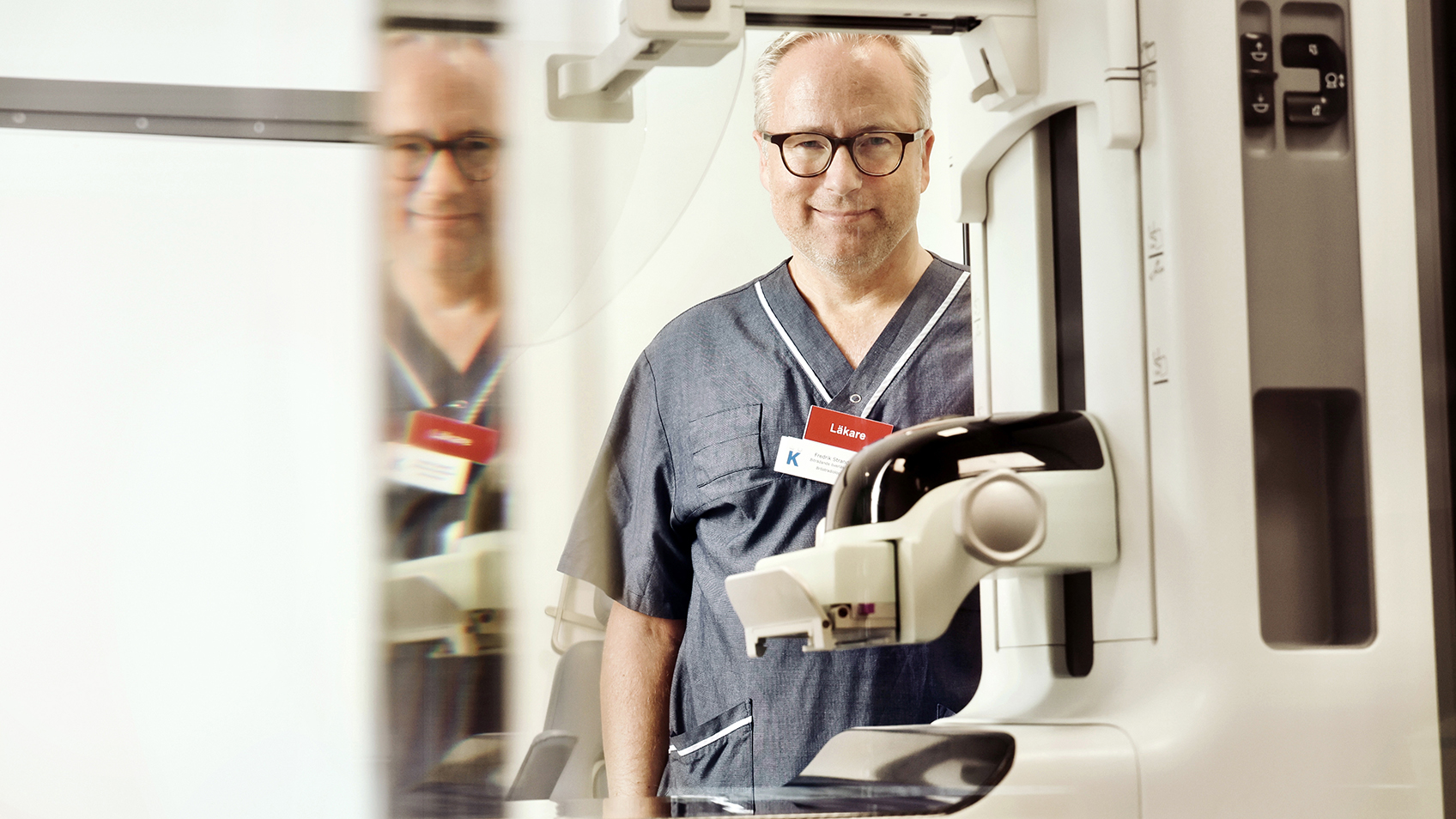
How do you see the future of artificial intelligence in breast cancer radiology?
-I think we were all surprised by chatGTP and how quickly it progressed. I think artificial intelligence in image diagnosis will also surprise us. The big difference is that chatGTP trains entirely online. We are limited by the fact that it is difficult to access the sensitive data we need. Therefore, it will take longer before we see the same significant progress here as in fields using publicly available data, says Fredrik Strand.
How good are AI algorithms?
– very good. At least as good as radiologists. But training the algorithm is much faster. In addition, algorithms can do things that radiologists are not trained to do, such as determine a patient’s risk of developing breast cancer in the future. The human brain also has a limited size, while artificial intelligence does not. While doctors, because of this limitation, have different specialties, AI will likely be good at everything, says Fredrik Strand.
Frederick Strand
age: 52 years old
Title: Doctor specializing in imaging and functional medicine at Karolinska University Hospital in Solna. Lecturer at Karolinska Institutet.
search: How AI can be used to evaluate radiological images from mammograms and MRIs.
Biggest research moment: When we actually saw that artificial intelligence contributed to increasing cancer detection.
Do in your free time: Reading, cooking for children, listening to P1.

“Extreme tv maven. Beer fanatic. Friendly bacon fan. Communicator. Wannabe travel expert.”



More Stories
Why Rare Earth Metals for Electric Cars Are Crucial for Modern Mobility
“We want to promote critical rules approach”
“A lot happened during the trip,” Jönköping County Council